By Julie H. Walker
Most of us associate fungus with itchy feet or hairy leftovers or, in the case of slime molds, the occasional unscrupulous lawyer. But to botany professor Wilford Hess, there is more to fungi than these common plagues that lap at the edge of shower doors, spot leaves in rose gardens, or put the bleu in cheese.
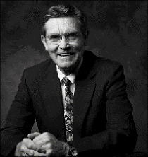
Wilford M. Hess, a professor of botany and range science, looks at fungus and other small parts of our big world a bit differently than most people, thanks to BYU’s microscopes.
As the director ofBYU’s microscopy lab and an expert on fungal spore morphology, Hess knows members of the mold family–the fungi, lichens, smuts, and rusts–at a highly intimate level. In his view the interior of a fungal spore is a fascinating architecture where tiny fungal factories crank out everything from mushrooms to medicines.
“Most people don’t realize just how much exposure we have to fungi and how they impact our daily lives. I’m fascinated by these little structures that have such an amazing ability to survive and adapt to new conditions,” he says.
Unlike plants, fungi do not contain chlorophyll and need only nutrients to grow. While fungal spores are microscopic–for example, 100 wheat smut spores can fit across the head of a pin–their fruiting structures, such as mushrooms and truffles, can be quite large. There are more than 100,000 species of fungi, including ancient specimens found in prehistoric fossilized plants.
Because their nutrition needs are diverse and they can survive in the harshest of conditions, fungi can be found on practically every square foot of the earth’s surface–from city stoops to forest floors.
Hess has studied many varieties of fungi, including many types that are important to human health and well-being. His current emphasis is endophytic fungi–those that live on or within plants. A single tree may have hundreds of species of endophytic fungi in its leaves, bark, or branches, but Hess says we know very little about their medicinal value.
“This is a field that a lot of people could work on for a lot of years and still they would only scratch the surface. In the tropics, you have very large numbers of endophytic fungi. One researcher said that in two species of trees in Central America, they isolated 600 species of fungi just from the leaves,” says Hess.
“Many, many kinds of antibiotics are produced by fungi, but it is only recently that we have begun to recognize endophytic fungi for their medicinal potential,” he says.
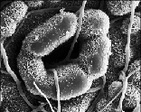
Fungus hyphal cells entering a stomate on a yew tree leaf (magnified 1,450 times)
Hess is currently involved in a project to investigate whether endophytic fungi can be used to produce a promising cancer treatment called taxol, which is found in species of yew trees. Because it is not found in high concentrations, and yew trees are rare, taxol costs $6,000 per gram. Hess’s research colleague, Gary Strobel of the University of Montana, determined that taxol could also be produced by fungi.
“Currently, the most important source of taxol is needles from Pacific yew trees. Large acreages are grown and harvested. We know that the fungi can take up the genetic capabilities to produce taxol. If taxol could be obtained from fungi, it would be much less expensive and a much better procedure environmentally. You could grow fungi in 50,000 gallon vats,” says Hess.
In December, Strobel and Hess published an extensive reference guide to leaves from species of yew trees, which includes Hess’s micrographs and descriptions of leaf patterns.
In addition to studying fungi with the potential to heal, Hess has conducted extensive studies on destructive fungal diseases, particularly smuts that affect wheat.
“In the United States alone, plant diseases cause $9.1 billion in crop losses annually. We need to know all we can about fungi so we can better understand how to more effectively
Cross section of a peccary hair (magnified 130 times)
control these devastating fungal diseases.”
He is also studying karnal bunt (KB) and other smut diseases that resist chemical treatment and have a devastating economic impact for American wheat farmers.
“KB causes a disease on our number one world food crop. We want to know how the spores function because it may help us to get a better insight into what we can do to help reduce the likelihood of disease,” says Hess.
Although karnal bunt spores are edible, large amounts can darken wheat kernels and change the taste of wheat flour. U.S. and foreign governments have aggressively policed the import and export of diseased wheat to stop its spread worldwide. While boxcars of valuable wheat sit in quarantine, American wheat producers lose money as they try to prove that the wheat is KB-free.
“The real problem is the quarantine costs. They’re not able to market their wheat where they want to because of the quarantines. The fungus is upsetting economics much more than it is upsetting biology. It is a real problem economically.”
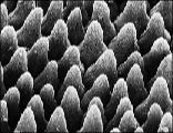
Cells on a snapdragon flower petal (magnified 250 times)
In addition to developing tests to identify bunt fungi, Hess is studying how bunt teliospores are so resistant to chemicals. A resilient layer within the spore wall is a source of frustration for those who hope to eradicate the disease. This same mysterious structure keeps fungal spores viable and allows them to lie dormant for years before germinating.
“There’s something chemically about that layer which makes it possible to set these little fungal spores aside and leave them for many years and then they’ll still germinate to retain the viability. I’d like to know the chemical nature of this layer and how it can keep the spores viable. I just wake up at night trying to figure out how to solve that problem–it’s absolutely fascinating,” says Hess.
In his 35 years on the BYU faculty, Hess has had his share of sleepless nights and long hours in the lab considering such scientific puzzles. He has always been a prolific researcher, maintaining high profile collaborations even while serving nine years as a department chair. In addition to teaching a full class load each semester and building a high-tech microscopy lab, he has produced more than 200 published works. His contributions to the university and his field have led to recognition with nearly every major university award for research and creative work, including the 1997 Karl G. Maeser Distinguished Fac-ulty Lecturer, the university’s top faculty honor.
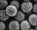
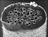
Wheat smut teliospores (magnified 1,450 times)
While much of his work has involved fungi, Hess has also studied other uses of microscopy. He established protocols for identifying and analyzing hair samples, information that can be used, for example, to create a criminal profile from a strand of hair or to assess exposure to drugs or chemicals.
Under his direction, the microscopy lab has been involved in projects from authenticating paint pigments for museums to studying the structural integrity of dental instruments to analyzing the content ofMark Hoffman’s ink samples. In addition to elemental analysis equipment, the BYU lab has an arsenal of high-powered microscopes–including a laser-scanning light microscope and scanning and transmission electron microscopes–that can magnify a specimen by as much as 200,000 times. Hess says these technologies change our view and increase our understanding in many ways.
“At each level of magnification, you see things that you don’t see at another level–it helps to understand how the biological and physical world is structured and how it functions,” Hess says.
So whether the subject of his study is the complex zig-zag texture that weaves across a fungal spore, the otherworldly craters on the surface of a cell, or the cathedral-window pattern that lies obscured in the cross section of a hair, Hess finds the microscopic world to be a constant source of fascination.
“Every time you use an electron microscope, you potentially see something that no one else has ever seen,” he says.
But Hess says that perhaps the most rewarding aspect of this work is that it leads to a greater appreciation of the deliberate precision and complexity of our world. For a scientist who is also a believer, every detail serves to be inspiring and faith affirming.
“I truly believe that when we see the very small, we see the hand of God. The more we look, the more complexity we see and the more it reveals the hand of God in all things.”