Cells are the building blocks of life—and one of the best ways to understand them, says Grant J. Jensen (BS ’94), dean of BYU’s College of Physical and Mathematical Sciences, “is to take terrific pictures of what’s inside.” With his research in cryotomography, Jensen has pioneered a new way to do just that.
Jensen and his research team at Caltech (where he taught before BYU) freeze cells to 80 Kelvin (that’s -292 Fahrenheit!), place them in a high-vacuum electron microscope, and photograph each sample as it slowly rotates. The photos are combined to create 3D models.
The resulting pictures revealed never-before-seen features of bacteria cells that have aided in understanding of how cells work. “Science is a wall,” Jensen says of his work. “Each discovery is one little brick in that wall.”
Going Places
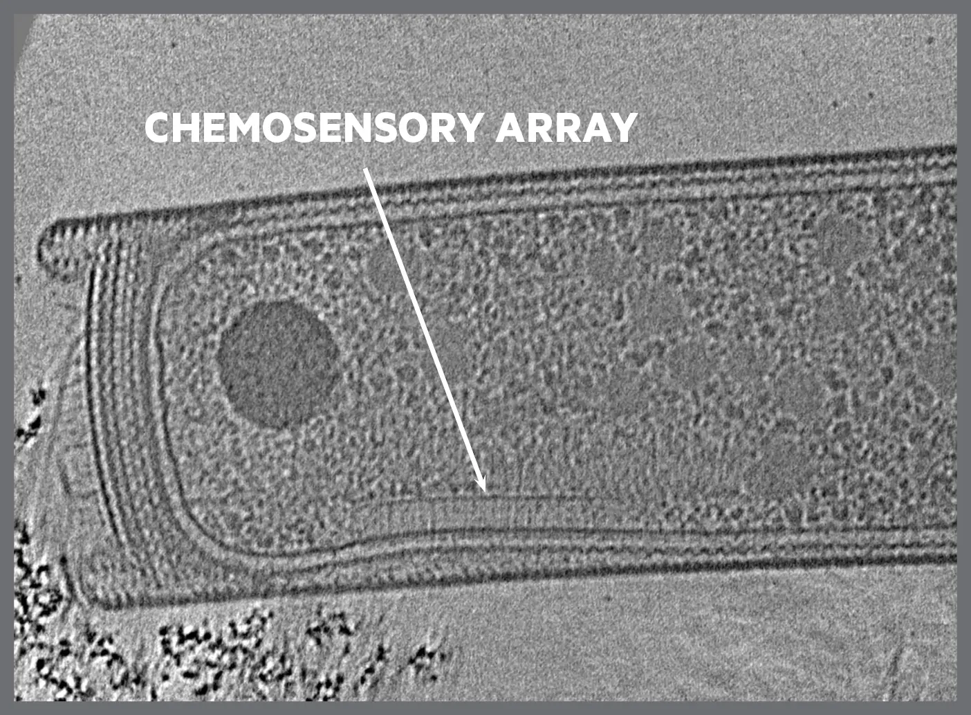
Chemosensory arrays are organelles that act as a “super nose,” helping cells sense chemical cues in the environment and move accordingly. Jensen’s team was able to see these structures for the first time, revealing a tightly knit network of sensitive interlocked receptors arranged in a honeycomb shape.
On the Offense
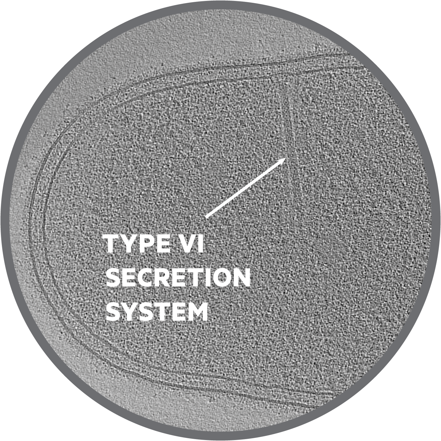
Survival is everything for cells, so offensive strategies are a must. Jensen and his team discovered a new one: the type VI secretion system. Or, as Jensen enthusiastically puts it, a “javelin thrower.” Cells with this system fire off a poison-tipped javelin that “kills the competitor by punching a hole in it,” Jensen says. “But sometimes it kills by depositing the toxin inside of the other cell. And it’s awesome!”
Lego Fortress
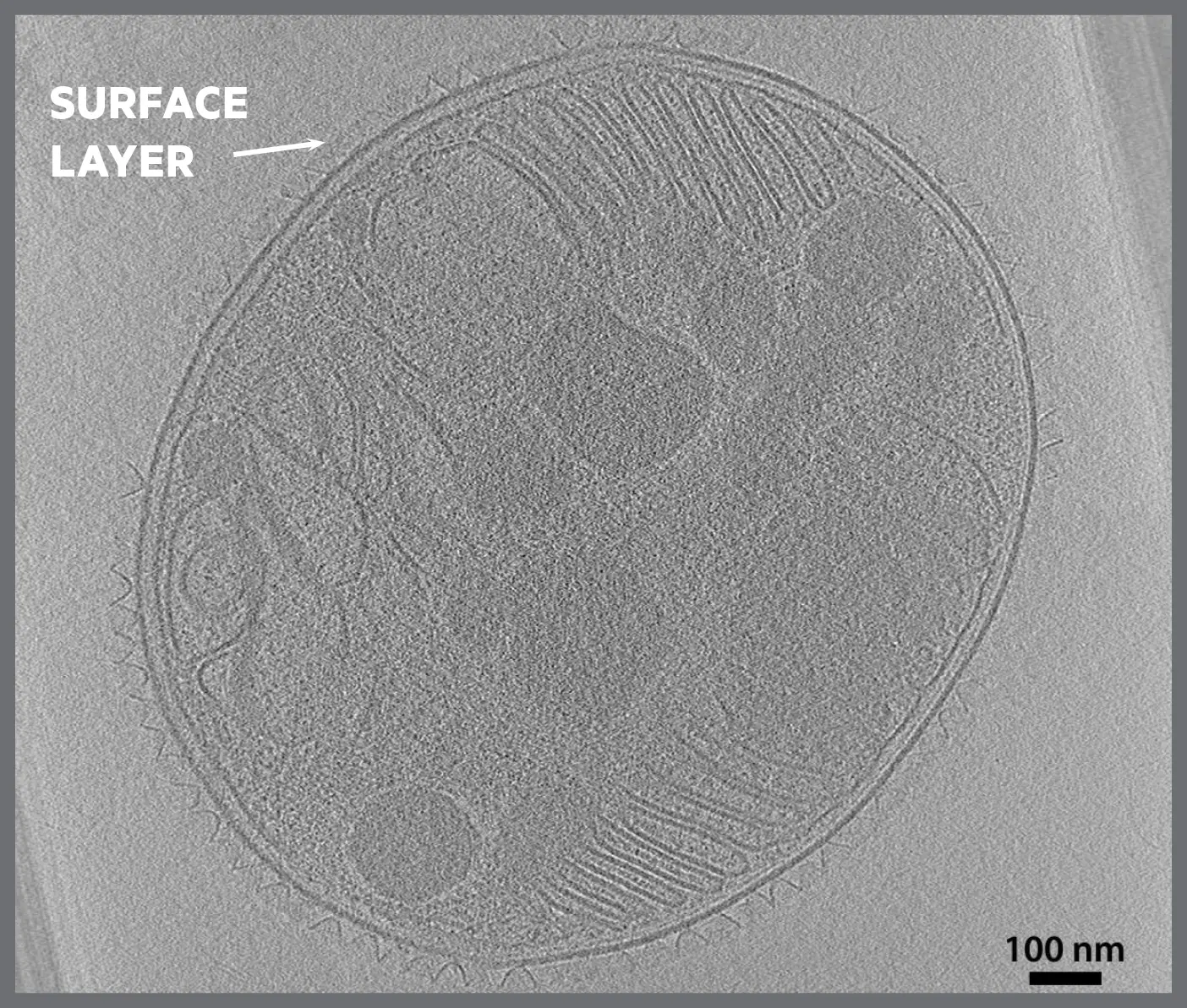
Some cells have protective surface layers that let nutrients in while blocking invaders. Cryotomography images have revealed that these are structured like Legos: cell-made proteins can pop in and out as the cell grows and changes. The appearance of the surface layer is different based on the proteins comprising it, which means there’s a lot more variability than was previously thought.