


Visualizing surgery is now a virtual reality.
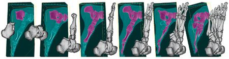
Live Surface allows surgeons to instantly visualize any part of a patient’s anatomy by extracting a 3-D computer image with just a few clicks of the mouse. THis sequence of images shows how the software can painlessly expose the bones of a foot.
A new software tool developed by BYU computer scientists will allow surgeons to quickly visualize any part of a patient’s anatomy by extracting a 3-D computer image from an MRI, CT scan, or 3-D ultrasounds with just a few clicks of the mouse.
The tool, dubbed “Live Surface” by its creators, professor William A. Barrett and graduate student Christopher J. Armstrong (BS ’04), also has special-effects applications—it can be used to extract inanimate objects or a single actor’s performance from video clips.
“The main goal in developing Live Surface was to give the physician a powerful, practical tool that can be used interactively,” says Barrett. Existing software and techniques used to give doctors a look at a patient’s anatomy are either too simplistic or take too long to be immediately useful. “A program like this has to be incredibly fast and very interactive or else it’s very frustrating for the user, who has to go get a sandwich and come back before he has what he wants.”
A breakthrough with Live Surface, says Barrett, is the ability to easily isolate “tricky” anatomy such as soft tissue—blood vessels, hearts, and muscles—which a lot of other techniques can’t readily extract.
All it takes is a click and drag of the mouse to isolate and extract the desired object. “This is the object I want, and this is not the object I want. And in less than half a second, it pulls the object you want out of the data,” says Armstrong, who presented the computer science research behind Live Surface at the International Workshop on Volume Graphics in July in Boston.
The program gets its speed from a hierarchical algorithm, or set of mathematical rules, that tells the computer to eliminate irrelevant information in broad, coarse cuts. Once the bulk of unwanted data is gone, the computer makes more refined calculations.
“It’s how you might envision cutting a toothpick out of a redwood tree,” says Armstrong. “You’d start with a chain saw and make very big cuts at first. By the end you’d use a knife to delicately shape the toothpick. Instead of ‘cuts’ our program intelligently identifies shapes. As you refine more and more, that shape becomes more exact.”
Barrett and Armstrong can think of an array of potential medical applications. Doctors could use the tool to make better diagnoses or more accurately locate cancerous tumors. Or after a surgeon has extracted a 3-D image of a person’s heart or brain, the image could then be projected onto the patient’s body, creating a road map for the surgeon as he operates.
Research for the software was partially funded by Adobe, makers of the popular image-editing program Photoshop. Barrett’s lab has had a long-running relationship with Adobe—Live Surface builds on Barrett’s development of Intelligent Scissors, known to Photoshop users as “magnetic lasso,” a program that allows users to quickly pull 2-D objects out of images.
Barrett and Armstrong hope Live Surface will become similarly ubiquitous in medical fields and make 3-D views of internal structures just a few clicks away for doctors and patients. “It’s really that simple,” says Armstrong.